HD PFM
HD PFM mode working principle also based on the “time window” approach.
In the user-defined “time window” referred to the tip-sample contact the AC voltage applied between conductive coating of the tip and investigated object.
AC voltage causes mechanical oscillations of the sample depending on its local polarization. Corresponding vertical (DFL siganl) and lateral (LF signal) motion of AFM tip is recorded in defined “time window” and processed to get amplitude and phase signals.
Amplitude of DFL and LF signals characterize local piezoelectric coefficient of the material whereas the phase signals give information about local polarization direction. The full DFL(t) curve of each HybriD mode circle is also processed to calculate adhesion, E modulus and feedback input signals. Thus HD PFM gives complex information about sample’s properties through a single measurement cycle, and at the same time, makes HD PFM nondestructive by retracting the tip from the sample at each HybriD oscillating cycle.
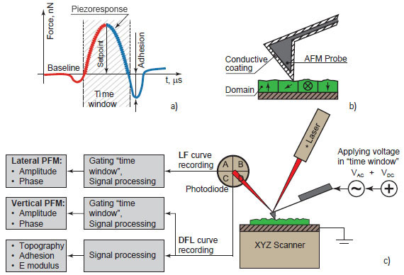
Figure 1. Working principle of HD PFM.
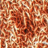
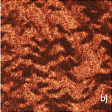
RESULTS AND DISCUSSION. COLLAGEN MATRIX
One of the most interesting biomaterials demonstrating piezoelectricity is collagen fibrils – the main building component of bones, teeth, corneal stroma and blood vessels. Collagen is composed of aligned polar protein molecules (fibrils) [21] that form a strong organic crystalline matrix. Piezoelectric properties of single collagen fibrils were recently studied with nanometer-level resolution [22,23]. Despite this advance, the scientific challenge of any piezoresponse study of collagen matrix is still tricky.
The main issue for traditional contact PFM measurements of these structures is complicated by rough surfaces where there is height variation of almost one micrometer. This makes AFM tip to cling to the single fibrils, and vice versa, highly distorting the topography and piezoresponse images.
Due to its working principle, HD PFM mode was found to be a superior technique for studying collagen matrix.
The samples were provided by FibraliSgn Corporation, where a unique technique of depositing animal collagen on glass substrate was developed [24].